

These videos are created to bridge the gap between theory and real-world surgical practice. Each case offers practical insights into decision-making, surgical techniques, anatomical understanding, and intraoperative challenges that are often not covered in textbooks. By observing step-by-step procedures and variations across different cases, gynecologists can refine their skills, improve precision, and build confidence in performing advanced laparoscopic surgeries.
The aim is to create a valuable learning resource where surgeons can continuously upgrade their knowledge!
Introducing a groundbreaking advancement in laparoscopic cerclage by Dr. Hrishikesh Pandit, Pandit Hospital, Ahilyanagar. This award-winning surgical technique presents a newer, safer, and more patient-friendly method of Laparoscopic Cerclage with a unique easy removal system that can be done through the vagina without opening the Pouch of Douglas and without any anesthesia. What Makes This Technique Revolutionary? Allows vaginal birth while retaining all advantages of laparoscopic cerclage Easy, painless stitch removal vaginally—no anesthesia required Reduces complications such as: Tape migration Vesicovaginal fistula Improved maternal comfort and safety
4 things can go wrong 1 Inadvertent ureteric injury Can be prevented by dissection close to vessel 2 Bladder injury Can be prevented by recognizing proper plane between cervix and bladder by identifying vertical capillaries and vascularity of plane 3 Vascular injury that is injury to uterine artery or vein That can be prevented by skeletanisation of uterine vessels . If bleeding is there after percising through tissue don’t get panic it may stop when you tie the knot of tape In worst scenario if uterine vessels get puncture you can sacrifice one sided vessel by coagulation it 4 Amniotic sac puncture This you can avoid it by making proper angel of the needle so that when you are turning the needle from posterior to anterior it should not go inside the cervical canal . For this you can pierce the needle close to the vesse
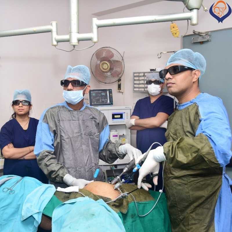
This video showcases a rare and challenging case of laparoscopic cervical cerclage in a patient with a pin-point cervix and absent vaginal portion of the cervix, where conventional vaginal cerclage was not possible. The patient had a history of multiple hysteroscopies for embryo transfer, resulting in complete cervical stenosis. The cervix was carefully dilated and intracervical fibrosis was released, following which embryo transfer became possible. A successful pregnancy was achieved, and cerclage was performed at 16 weeks of gestation. Key highlights of this case: Absent vaginal cervix, necessitating the use of alternative anatomical landmarks Uterine artery used as a landmark, with the tape passed medial to the uterine vessels Careful dissection and skeletonisation of uterine vessels Creation of space between the uterus and vessels Tape passed from anterior to posterior without piercing uterine tissue Uterine approach avoided due to ballooned lower uterine segment and high risk of amniotic sac puncture Procedure performed in a unicornuate uterus, adding to the complexity This video demonstrates meticulous surgical planning, advanced laparoscopic skills, and highlights the importance of anatomical understanding in complex fertility-preserving procedures. Enjoy the visualisation of the pregnant uterus and uterine vessel skeletonisation.
👉🏼First Time of YouTube Presenting a rare and challenging case of laparoscopic cervical cerclage in a pregnant patient with a history of: Previous LSCS (Lower Segment Cesarean Section) Two failed vaginal cerclages Two second-trimester pregnancy losses Key Surgical Challenges & Insights:
✅Dense Bladder Adhesions: Due to prior cesarean section, there is significant bladder adhesion. Observe how the assistant provides counter-traction to help in dissection and exposure.
✅Fibrosis Around Right Uterine Artery The right uterine artery is pulled up with surrounding fibrosis. Careful creation of a window in the broad ligament is essential—watch how the dissection stays close to the round ligament to avoid vascular injury.
✅Engorged Veins on Left Broad Ligament Increased vascularity adds to the complexity. Watch the meticulous handling of the area to prevent bleeding and ensure patient safety. This video is an excellent educational resource for gynecologists and surgeons interested in advanced laparoscopic techniques.
This video is an excellent educational resource for gynecologists and surgeons interested in advanced laparoscopic techniques.
I provide, laparoscopy surgery training for gynecologists who wants to master the techniques of laparoscopy surgery.
– Dr. Hrishikesh Pandit (Laparoscopic Gynecology Surgeon)
In the last 5 years of training, Dr. Hrishikesh Pandit has trained more than 500 gynecology surgeons from across India and other countries as well (United Kingdom, United Arab Emirates and Kenya etc.) As he is working with many leading gynecological surgery hospitals in Ahmednagar district and other parts of Maharashtra, he has a continuous flow of patients who require laparoscopic surgical treatment. The volume of patients provide advantage for all the trainees to practice hands-on laparoscopy technique under the guidance of Dr. Hrishikesh Pandit.
Dr. Hrishikesh Pandit : Best Laparoscopic Surgeon | LET’S SEE OUR INTRO VIDEO
FAQ
You should consult a doctor during the first 6 to 8 weeks of your pregnancy, or when your period is 2 to 4 weeks late.
If your contractions are 5 minutes apart, lasting for 1 minute, for 1 hour or longer, it’s time to head to the hospital.
Doctors recommend an infertility evaluation if you have not gotten pregnant after 1 year of having regular sexual intercourse without using birth control. If you are older than 35, an evaluation is recommended after 6 months of trying.
Yes, You can. But most babies need 39 weeks to develop fully. Induced or planned delivery before that time—without a valid medical reason—is not in the best interest of the baby or the mother. After 39 weeks you can plan delivery.
Women who are 21 to 29 should have a Pap test alone every 3 years. HPV testing alone can be considered for women who are 25 to 29, but Pap tests are preferred. Women who are 30 to 65 have three options for testing. They can have a Pap test and an HPV test (co-testing) every 5 years. They can have a Pap test alone every 3 years. Or they can have HPV testing alone every 5 years.
Laparoscopic hysterectomy is a safe and suitable procedure for chosen patients. It affords patients advantages like less peri-operative morbidity, better life quality, shorter hospitalization time, and faster return to activity.
Schedule a doctor’s visit if you have: Greenish, yellowish, thick or cheesy vaginal discharge; Strong vaginal odor; Redness, itching, burning or irritation of your vagina or the area of skin that surrounds the vagina and urethra (vulva); Bleeding or spotting unrelated to your period.
Painless delivery can be achieved using a form of regional anesthesia that provides pain relief during natural labor. Epidural anesthesia is administered through an injection on the lower back of the mother. The drug takes about 10-15 minutes to take effect.
Even in severe cases of endometriosis, most can be treated with laparoscopic surgery. In laparoscopic surgery, your surgeon inserts a slender viewing instrument (laparoscope) through a small incision near your navel and inserts instruments to remove endometrial tissue through another small incision.
The HPV vaccine is recommended for routine vaccination at the age of 11 or 12 years. (Vaccination can be started at age 9.) It is also recommended that vaccination for everyone through age 26 years if not adequately vaccinated when younger. HPV vaccination is given as a series of either two or three doses, depending on age at initial vaccination.